
|
Upload | Sign in |
|---|
Medical imaging is the visualization of body parts, tissues, or organs, for use in clinical diagnosis, treatment and disease monitoring. Imaging techniques encompass the fields of radiology, nuclear medicine and optical imaging and image-guided intervention.
It refers to the refers to several different technologies that are used to view the human body in order to diagnose, monitor, or treat medical conditions. Each type of technology gives different information about the area of the body being studied or treated, related to possible disease, injury, or the effectiveness of medical treatment.
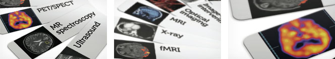
The various Medical Imaging techniques are:-
Ultrasound Imaging- uses sound waves to produce images of the interior of the body.
MRI (Magnetic Resonance Imaging)- uses magnetic fields rather than X-rays to produce 3-D images.
Radiography - Uses radiation to produce 2-D images (x-rays) or 3-D scans (CT). 3- images can be manipulated with software to provide views from any angle.
Computed Tomography (CT)- Computed tomography (CT scanning) is a medical imaging modality where tomographic images or slices of specific areas of the body are obtained from a large series of two-dimensional X-ray images taken in different directions.These cross-sectional images can be combined into a three-dimensional image of the inside of the body and used for diagnostic and therapeutic purposes in various medical disciplines.
Fluoroscopy - Fluoroscopy is an imaging technique commonly used by physicians or radiation therapists to obtain real-time moving images of the internal structures of a patient through the use of a fluoroscope. In its simplest form, a fluoroscope consists of an X-ray source and fluorescent screen between which a patient is placed. However, modern fluoroscopes couple the screen to an X-ray image intensifier and CCD video camera allowing the images to be recorded and played on a monitor. This method may use a contrast material.For Ex:cardiac catheterization to examine coronary artery blockages
Mammography-Mammography is the process of using low-energy X-rays (usually around 30 kVp) to examine the human breast and is used as a diagnostic and a screening tool. The goal of mammography is the early detection of breast cancer, typically through detection of characteristic masses and/or microcalcifications.
Why is medical imaging important?
In an ideal world we would be able to diagnose, treat and cure patients without causing any harmful side effects. The use of medical imaging has enabled doctors to see inside a patient without having to cut them open. Medical imaging also helps us learn more about neurobiology and human behaviours. For brain imaging is being used to understand why some people become long-term cocaine addicts and some do not. Medical imaging brings scientists from biology, chemistry and physics together and the technologies developed can often be used in many disciplines.
blog comments powered by Disqus