
|
Upload | Sign in |
|---|
Magnetic Resonance Imaging (MRI)
Magnetic resonance imaging is a medical imaging technique used in radiology to visualize internal structures of the body in detail.
The principle behind the use of MRI machines is that they make use of the fact that body tissue contains lots of water (and hence protons) which gets aligned in a large magnetic field. MRI uses a powerful magnetic field, radio frequency pulses and a computer to produce detailed pictures of organs, soft tissues, bone and virtually all other internal body structures.
The images can then be examined on a computer monitor, transmitted electronically, printed. MRI does not use ionizing radiation (x-rays).
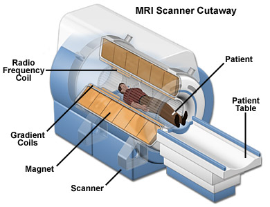
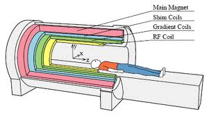
An MRI scanner is a device in which the patient lies within a large, powerful magnet where the magnetic field is used to align the magnetization of some atomic nuclei in the body, and radio frequency fields to systematically alter the alignment of this magnetization.
This causes the nuclei to produce a rotating magnetic field detectable by the scanner—and this information is recorded to construct an image of the scanned area of the body.
The basis of MRI (Magnetic Resonance imaging) is the directional magnetic field, or moment, associated with charged particles in motion.
Nuclei containing an odd number of protons and/or neutrons have a characteristic motion or precession. Because nuclei are charged particles, this precession produces a small magnetic moment.
When a human body is placed in a large magnetic field, many of the free hydrogen nuclei align themselves with the direction of the magnetic field.
The nuclei precess about the magnetic field direction like gyroscopes.
This behaviour is termed Larmor precession.
The frequency of Larmor precession is proportional to the applied magnetic field strength as defined by the Larmor frequency : ω0=γB0………… (1) Where γ is the gyrometric ratio and B0 is the strength of applied magnetic field.
For hydrogen, Gyrometric ratio = 42.6 MHz / Tesla.
To obtain an MR image of an object, the object is placed in a uniform magnetic field, B0 , of between 0.5 to 1.5 Tesla. As a result, the object's hydrogen nuclei align with the magnetic field and create a net magnetic moment, M, parallel to B0 .
This behaviour is illustrated in following fig. 1:
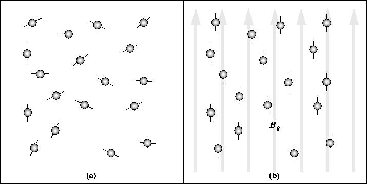
In the absence of a strong magnetic field, hydrogen nuclei are randomly aligned as in (a). When the strong magnetic field, B0, is applied, the hydrogen nuclei precess about the direction of the field as in (b).
Next, a radio-frequency (RF) pulse, Brf , is applied perpendicular to B0. This pulse, with a frequency equal to the Larmor frequency, causes to M tilt away from B0 as in following fig.2.
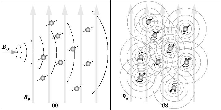
The RF pulse, Brf , causes the net magnetic moment of the nuclei, M, to tilt away from B0 . (b) When the RF pulse stops, the nuclei return to equilibrium such that M is again parallel to B0. During realignment, the nuclei lose energy and a measurable RF signal is emitted.
This return to equilibrium is referred to as relaxation.
During relaxation, the nuclei lose energy by emitting their own RF signal (see Figure 2.b). This signal is referred to as the free-induction decay (FID) response signal.
The FID response signal is measured by a conductive field coil placed around the object being imaged. This measurement is processed or reconstructed to obtain 3D grey-scale MR images.
To produce a 3D image, the FID resonance signal must be encoded for each dimension. The encoding in the axial direction, the direction of B_0, is accomplished by adding a gradient magnetic field to B0.
This gradient causes the Larmor frequency to change linearly in the axial direction. Thus, an axial slice can be selected by choosing the frequency of Brf to correspond to the Larmor frequency of that slice.
The 2D spatial reconstruction in each axial slice is accomplished using frequency and phase encoding. A ``preparation'' gradient, Gy, is applied causing the resonant frequencies of the nuclei to vary according to their position in the Y-direction.
Gy is then removed and another gradient, Gx, is applied perpendicular to Gy.
As a result, the resonant frequencies of the nuclei vary in the X-direction due to Gx and have a phase variation in the Y-direction due to the previously applied Gy.
Thus, X-direction samples are encoded by frequency and Y-direction samples are encoded by phase.
A 2D Fourier Transform is then used to transform the encoded image to the spatial domain.
The voxel intensity of a given tissue type (i.e. white matter vs. grey matter) depends on the proton density of the tissue; the higher the proton density, the stronger the FID response signal. MR image contrast also depends on two other tissue-specific parameters:
1) The longitudinal relaxation time, T1, and
2) The transverse relaxation time, T2.
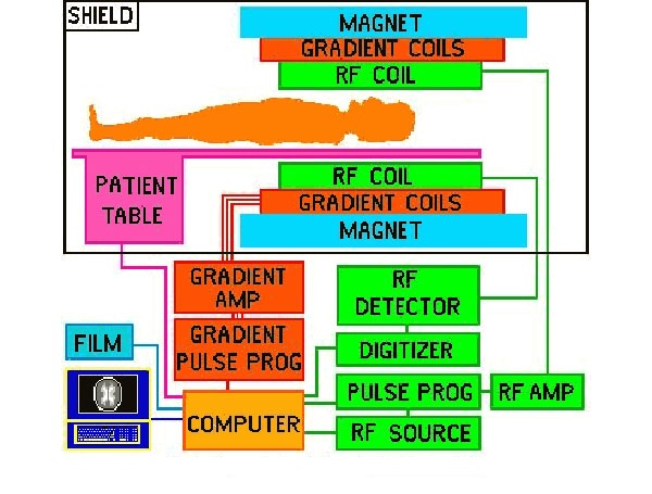
Block diagram:
Other than MRI there is a another concept called Functional MRI;
Functional Magnetic Resonance Imaging or FMRI is a non-invasive technique for imaging the activation of brain areas by different types of physical sensation (sight, sound, touch, taste, smell) or activity such as problem solving and/or movement (limited by the machine).
Thus, FMRI scans are an increasingly common tool for “brain mapping” in cognitive science.
FMRI scans use the same basic principles of atomic physics as MRI scans, but MRI scans image anatomical structure whereas FMRI image metabolic function. Thus, the images generated by MRI scans are like three dimensional pictures of anatomic structure. The images generated by FMRI scans are images of metabolic activity within these anatomic structures