
|
Upload | Sign in |
|---|
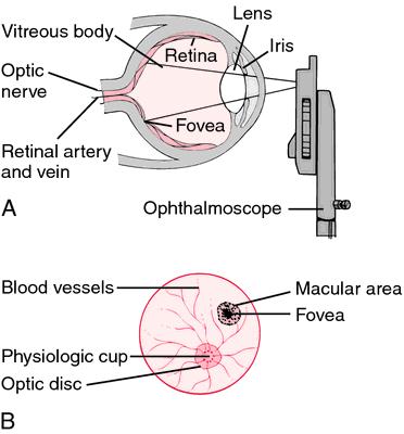
Ophthalmoscopy (funduscopy or fundoscopy) is a test that allows a health professional to see inside the fundus of the eye and other structures using an ophthalmoscope (or funduscope).
It is done as part of an eye examination and may be done as part of a routine physical examination. It is crucial in determining the health of the retina and the vitreous humor.
It is of two major types:
• Direct ophthalmoscopy one that produces an upright, or unreversed, image of approximately 15 times magnification.
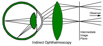
• Indirect ophthalmoscopy one that produces an inverted, or reversed, direct image of 2 to 5 times magnification.
Features |
Direct ophthalmoscopy |
Indirect ophthalmoscopy |
Condensing lens |
Not Required |
Required |
Examination distance |
As close to patient's eye as possible |
At an arm's length |
Image |
Real, inverted |
|
Illumination |
Not so bright; so not useful in hazy media |
Bright; so useful for hazy media |
Area of field in focus |
About 2 disc diameters |
About 8 disc diameters |
Stereopsis |
Absent |
Present |
Accessible fundus view |
Slightly beyond equator |
Up to Ora serrata i.e. peripheral retina |
Examination through hazy media |
Not possible |
Possible |
Each type of ophthalmoscopy has a special type of ophthalmoscope:
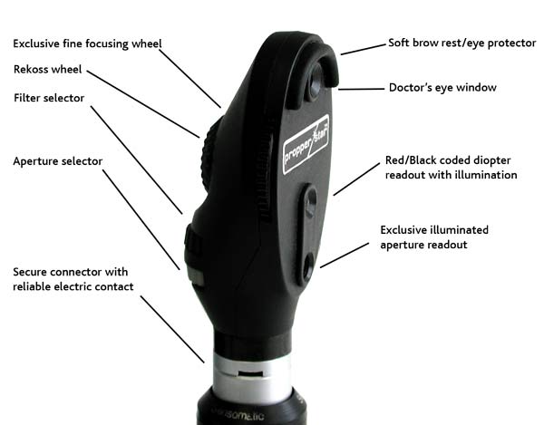
Direct ophthalmoscope one that produces an upright, or unreversed, image of approximately 15 times magnification.
The direct ophthalmoscope is used to inspect the fundus of the eye, which is the back portion of the interior eyeball. Examination is best carried out in a darkened room.
The examiner looks for changes in the colour or pigment of the fundus, changes in the caliber and shape of retinal blood vessels, and any abnormalities in the macula lutea, the portion of the retina that receives and analyzes light only from the very center of the visual field.
Macular degeneration and opacities of the lens can be seen through direct ophthalmoscopy.
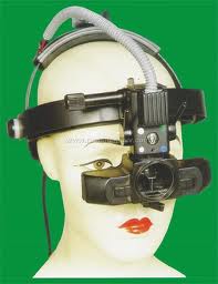
Indirect ophthalmoscope one that produces an inverted, or reversed, direct image of two to five times magnification.
An indirect ophthalmoscope provides a stronger light source, a specially designed objective lens, and opportunity for stereoscopic inspection of the interior of the eyeball.
It is invaluable for diagnosis and treatment of retinal tears, holes, and detachments.
The pupils must be fully dilated for satisfactory indirect ophthalmoscopy.
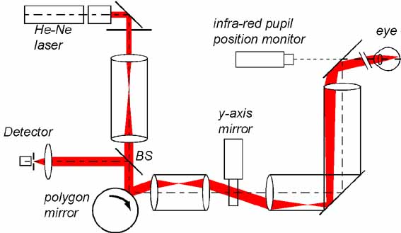
Scanning laser ophthalmoscope (SLO) an instrument for retinal imaging in which light from a low-power laser beam that scans the retina is reflected back to a sensor; the light detected by the sensor is used to create a full-colour composite digital image. Block diagram of SLO
blog comments powered by Disqus